All Elbow Fat Pads Are Best Demonstrated in Which Position
What structures are comprising the neural or verterbral arch. All elbow fat pads are best demonstrated in which position.

100 Top Mcdavid Dizlik Basketball Leg Sleeve Breathable Sports Honeycomb Pad Bumper Barce Kneelet Protectores Rodilleras De Voleibol Rodilleras Moda Deportiva
That a fat pad sign may demonstrate a false positive if the lateral has been improperly positioned.
. Elbow flexed 90 degrees. Therefore in a noninjured correctly positioned lateral elbow the posterior fat pad should not. Place the arm on the table with elbow straight.
Anteroposterior AP extended 21. What is clearly seen in profile. They are hyperechoic and triangular and are displaced with distention of the joint 45.
All elbow fat pads are best demonstrated in which position. The AP projection is made through the partially flexed elbow resting on the olecranon process CR parallel to the humerus. A small amount of normal fluid may be seen between the anterior fat pad and humerus.
With the patient seated at the end of the x-ray table elbow flexed 80 degrees and the CR directed 45 degrees laterally from the shoulder to the elbow joint which of the following structures will be demonstrated best. All elbow fat pads are best demonstrated in which position. Trauma or other pathology.
The tarsals and metatarsals are arranged to form the. Are either partially or completely superimposed on all projections of the elbow. Radial tuberosity facing anteriorly.
The elbow fat pads are situated external to the synovium and within the fibrous external joint capsule. All elbow fat pads are best demonstrated in which position. To visualize fat pads surrounding the elbow exposure factors must be adjusted to see both bony and soft tissue structures.
Both anterior and posterior fat pad signs exist and both can be found on the same X-ray. Gastric or bowel muscoa. 51 rows All elbow fat pads are best demonstrated in what position.
There is an intraarticular fracture of the radial head. Which position of the shoulder demonstrates the lesser tubercle in profile medially. Anterior and posterior fat pads of the elbow are best seen on correctly positioned and correctly exposed anteroposterior AP clbow projections.
Lateral Which surface must be adjacent to the IR to obtain a lateral projection of the fourth finger with optimal recorded detail. The posterior fat pad is not seen because it is located in the intercondylar fossa. 107the term used to describe the presence of blood in vomit is.
It is caused by displacement of the fat pad around the elbow joint. The following should be clearly demonstrated. All elbow fat pads are best demonstrated in which position.
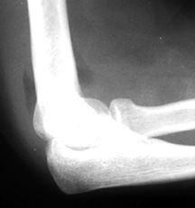
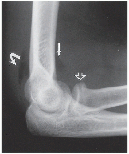
All elbow fat pads are best demonstrated in which position. Lateral radiograph shows elevated anterior and posterior fat pads indicative of joint effusion. Open elbow joint centered to the central ray.
When the elbow is flexed the posterior fat pad lies deep within the olecranon fossa and is hidden from view by the medial and lateral condyles. Double-contrast examinations of the stomach or large bowel are performed to better visualize the. All elbow fat pads are best demonstrated in which position.
Which of the following are fat pads or fat stripes that may be visible on the lateral projection of the elbow during trauma. All elbow fat pads are best demonstrated in which position. Double contrast exams of the stomach or large bowel are performed to better visualize the.
Normally when the elbow is flexed to 90 the anterior fat pad may be seen just anterior to the joint. Study free Radiology flashcards about RADT 465 Procedures created by 18Fieldsms to improve your grades. What projection to best to view the pisiform.
With joint distention the fat pads are displaced away from the joint in the anterior aspect. The number of phalanges in the thumb The patients chin should be elevated during chest radiography to All elbow fat pads are best demonstrated in this position The projectionmethod often used to detect carpal canal defect The trapezium articulates with this bone The capitulum articulates with this This position demonstrates the scaphoid. Trauma or other pathology.
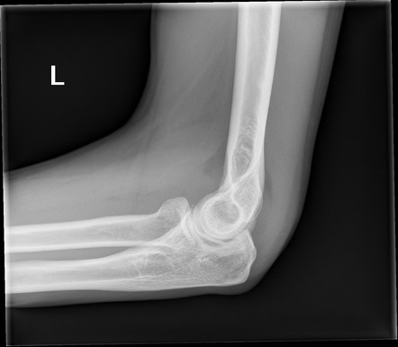
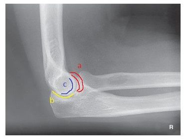
Other angles may tempt the posterior fat pad to emerge from the olecranon fossa mimicking the fat pad sign. Position of patient The patient should be Seated sideways at the end of the the table. Ideally the upper arm elbow and forearm are all resting on the table.
Demonstration of the posterior fat pad on the lateral projection of the adult elbow can be caused by 1. Which position of the shoulder demonstrates the lesser tubercle medially. 1 and 2 only.
All elbow fat pads are best demonstrated in which position. For the Coyle method elbow describe tube angle and patient position. Intracapsular extrasynovial elbow fat pads are found between the hypoechoic synovial lining and hyperechoic linear fibrous capsule within the fossae.
Pedicles and the Laminae. Proper positioning requires 90 flexion of the elbow joint. The posterior fat pad is located in the olecranon fossa and the anterior fat pad in the coronoid fossa.
Elbow Arm and Shoulder. Axial CT shows the fracture traversing the articular surface of the radial head. A Symphysis B Rami C Body D Angle.
The actual wrist is made up of which bones. The term used to describe the presence of blood in vomit. Matching game word search puzzle and hangman also available.
With the patients head in a PA position and the CR directed 20 degrees cephalad which part of the mandible will be best visualized. Radial head partially superimposing the coronoid process. The fat pad will be elevated away from the joint and the posterior fat pad will be visible.
Demonstration of the posterior fat pad on the lateral projection of the adult elbow can be caused by. The shoulder girdle is part of which skeletal system. Demonstration of the posterior fat pad on the lateral projection of the adult elbow can be caused by 1.
Gastric or bowel mucosa. All elbow fat pads are best demonstrated in a lateral postion. A AP B Lateral C Acute flexion D AP partial flexion.

The Pediatric Elbow A Review Of Fractures
Imaging Radial Head Fractures Wikiradiography
Fat Pad Signs In Elbow Trauma Document Gale Onefile Health And Medicine

Soft Tissue Signs The Elbow Body Anatomy Radiology Pediatrics

Pdf Rapid Screening For The Posterior Fat Pad Sign In Suspected Pediatric Elbow Fractures Using Point Of Care Ultrasound A Fast Exam For The Traumatized Elbow
Imaging Radial Head Fractures Wikiradiography

Posterior Fat Pad Sign Elbow Radiology Reference Article Radiopaedia Org

The Spectrum Of Elbow Fracture Patterns Is Shown Download Scientific Diagram

Comparison Lateral Elbow Radiographs In A Child Who Sustained A 5 Cm Download Scientific Diagram
How To Screen A Paediatric Elbow X Ray For Injuries

References In Radiography Of The Radial Head An Alternative View Radiography

Pdf Rapid Screening For The Posterior Fat Pad Sign In Suspected Pediatric Elbow Fractures Using Point Of Care Ultrasound A Fast Exam For The Traumatized Elbow

Pin On Radiology Human Xray Anatomy

Upper Limb Ii Elbow Radiology Key

Comparison Lateral Elbow Radiographs In A Child Who Sustained A 5 Cm Download Scientific Diagram

Pdf Rapid Screening For The Posterior Fat Pad Sign In Suspected Pediatric Elbow Fractures Using Point Of Care Ultrasound A Fast Exam For The Traumatized Elbow

Posterior Fat Pad Sign Elbow Radiology Reference Article Radiopaedia Org

Changing The Way You Learn Flashcards

References In A Pilot Study To Examine The Effect Of An Educational Poster On The Knowledge And Practices Of Lateral Elbow Radiograph Repositioning In Radiographers Journal Of Medical Imaging And Radiation
Comments
Post a Comment